.png)


AI-Powered Brain Atlas Maps the Human Mind with Unprecedented Detail
- Covertly AI
- Nov 9, 2025
- 3 min read


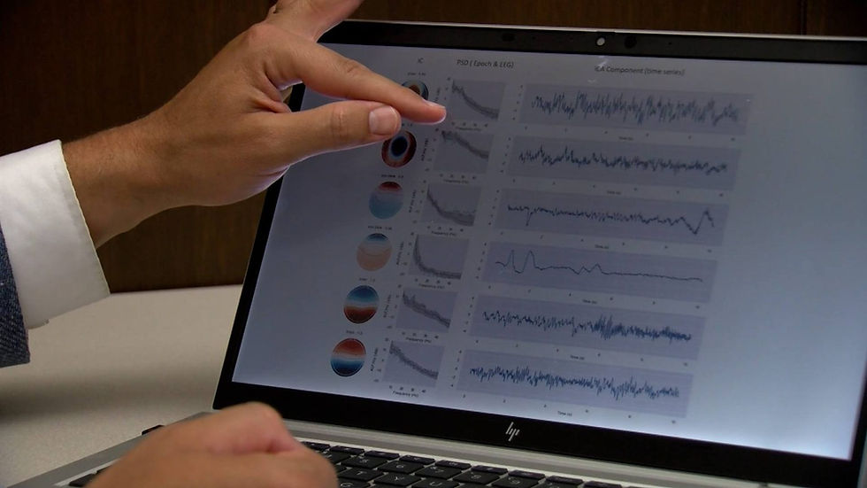
A team of researchers from University College London (UCL) has developed an advanced AI-assisted brain atlas that allows scientists to visualize the human brain in unprecedented detail, marking a major milestone in neuroscience and neuroimaging (News-Medical.net; Medical Xpress; Mirage News). Named NextBrain, the atlas provides a comprehensive map of the adult human brain, enabling the analysis of MRI scans in just minutes while offering a level of resolution that was previously unachievable.
The human brain is made up of hundreds of interconnected regions responsible for driving thought, emotion, and behavior. Traditional brain atlases have long been able to identify major structures such as the hippocampus, an area vital for memory and learning, but have struggled to distinguish their finer sub-regions. These distinctions are critical since different sub-regions can be affected unevenly in neurodegenerative conditions like Alzheimer’s disease (News-Medical.net). While microscopic imaging, or histology, can reveal cellular-level details, this process cannot be applied to living individuals, making it challenging to study how the brain evolves with age or disease.

Published in Nature, NextBrain bridges this gap by combining microscopic data with MRI imagery through the use of artificial intelligence. The result is a freely available atlas that researchers worldwide can use to examine the living brain with unprecedented precision. The project took six years to complete and involved dissecting, staining, and photographing 10,000 sections from five post-mortem human brains. Each brain was scanned with MRI before dissection to ensure accurate digital reconstruction, much like assembling a three-dimensional jigsaw puzzle with the original picture as a guide (Medical Xpress).
Artificial intelligence played a central role in aligning thousands of microscope images with MRI scans, compensating for differences in imaging techniques and ensuring seamless reconstruction. In total, 333 distinct brain regions were labeled within the five reconstructed models, a task that would have taken decades to complete manually without AI assistance. Dr. Juan Eugenio Iglesias, senior author of the study from UCL Medical Physics & Biomedical Engineering and Massachusetts General Hospital/Harvard Medical School, explained that NextBrain represents “the culmination of years of effort to bridge the gap between microscope imaging and MRI.” By merging high-resolution tissue data with AI techniques, the team created a tool that provides insights into neurodegenerative diseases and aging that were previously beyond reach (News-Medical.net).
The resulting atlas is an “average” model of the human brain that can be generalized across individuals, whether living or deceased. Its reliability was validated through thousands of MRI datasets taken under varying imaging conditions and using different scanner types. In one key test, the atlas accurately identified tiny brain structures, such as the subregions of the hippocampus, when labeling an ultra-high-resolution MRI scan. In another large-scale application, the research team analyzed more than 3,000 MRI scans of living participants to study age-related brain changes, achieving a level of anatomical detail that existing tools could not match (Medical Xpress).

According to Dr. Zane Jaunmuktane from UCL’s Queen Square Institute of Neurology and the Queen Square Brain Bank for Neurological Disorders, the team’s goal was to enable researchers to “identify hundreds of brain regions in living patients quickly and consistently, while maintaining the fine-grained anatomical accuracy of microscope data.” NextBrain offers an unparalleled view of the brain’s cellular architecture, enabling the early detection of neurological diseases such as Alzheimer’s before clinical symptoms arise (News-Medical.net; Mirage News).
All the data, tools, and annotations from NextBrain have been made openly accessible through the FreeSurfer neuroimaging platform, which also provides visualization and educational resources to facilitate global use. Supported by the European Research Council, the Alzheimer’s Society, the Lundbeck Foundation, and the U.S. National Institutes of Health, the development of NextBrain signals a transformative leap forward in brain mapping technology, one that promises to accelerate discoveries and improve the diagnosis and treatment of neurological disorders.
This article was written by the Covertly.AI team. Covertly.AI is a secure, anonymous AI chat that protects your privacy. Connect to advanced AI models without tracking, logging, or exposure of your data. Whether you’re an individual who values privacy or a business seeking enterprise-grade data protection, Covertly.AI helps you stay secure and anonymous when using AI. With Covertly.AI, you get seamless access to all popular large language models - without compromising your identity or data privacy.
Try Covertly.AI today for free at www.covertly.ai, or contact us to learn more about custom privacy and security solutions for your business.
Works Cited
“New AI-Assisted Atlas Can Help Visualize the Human Brain in Unprecedented Detail.” News-Medical.net, 5 Nov. 2025, https://www.news-medical.net/news/20251105/New-AI-assisted-atlas-tcan-help-visualize-the-human-brain-in-unprecedented-detail.aspx.
“New Brain Atlas Offers Unprecedented Detail in MRI Scans.” Medical Xpress, 5 Nov. 2025, https://medicalxpress.com/news/2025-11-brain-atlas-unprecedented-mri-scans.html.
“New Brain Atlas Reveals Unseen MRI Detail.” Mirage News, 5 Nov. 2025, https://www.miragenews.com/new-brain-atlas-reveals-unseen-mri-detail-1564580/.
Study.eu. “University College London, United Kingdom | Study at University College London.” Study.eu, 2025, https://www.study.eu/university/university-college-london.


Comments